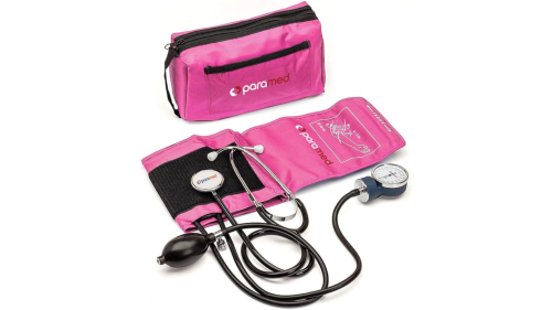
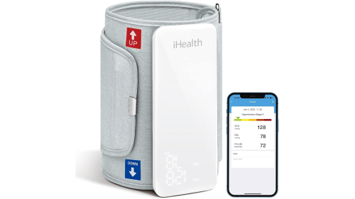
Vital signs are intended to give an EMS provider a picture of the current physiologic status of his patient. Most EMS practitioners learned vital signs early in their education and can sometimes forget how meaningful an accurate set of vitals can be. Many providers are taught that when they are the team leader to delegate vital signs and it simply becomes habit to ask another responder to obtain vitals and to take those result at face value.
Given that vitals are a snapshot of the patient’s physiologic status perhaps they deserve more attention and importance. Here are five ways to improve your capturing, monitoring and interpreting of a patient’s vitals.
1. Avoid assuming a systolic pressure based on a pulse location
Many EMS providers learned that a palpable pulse at a specific anatomic location correlates to an estimated systolic blood pressure. Most commonly, providers are taught that a radial pulse means a systolic of at least 90 mm Hg, a femoral pulse 70 mm Hg and a carotid pulse 60 mm Hg.
This assumption was historically taught in certification courses including Advanced Trauma Life Support, but is not supported by peer-reviewed research. The assessment work-around has since been pulled out of most standard curricula, but the practice continues in EMS, likely as one of the all-too-persistent traditions within medicine that hangs around because “we’ve always done it that way”.
While not a substitute for a complete blood pressure measurement, a present palpable pulse does inform the EMS provider of a few important conditions. First, a palpable pulse confirms that the patient has a heartbeat and some level of cardiac output. Additionally, the presence of a radial pulse can generally infer adequate perfusion to the brain. Finally, a comparison of pulse rate and quality between the left and right extremities can assist in identifying a vascular condition like an aortic aneurysm. What a palpable pulse cannot do is infer a systolic blood pressure measurement.
2. Take a full blood pressure
Along the same lines as estimating blood pressure based on the location of a palpated pulse, taking only a systolic blood pressure provides merely a piece of the intended measurement. Blood pressure is a measure of the pressure in the arteries during systole (the heart pumping) and diastole (the heart at rest).
Measuring only the systolic blood pressure — known as palpating a blood pressure — does give an idea of whether the organs of the body are being perfused with blood, but taking both systolic and diastolic pressure allows for the calculation of mean arterial pressure.
MAP can be calculated using a variety of equations but many cardiac monitors will display the value based on a measured blood pressure. MAP represents the pressure of blood perfusing the organs and, as such, gives a better estimate of how effective the circulatory system is functioning.
A MAP of 70 mm Hg is considered sufficient to perfuse the organs and a normal range for MAP is between 60 and 110 mm Hg. A falling MAP measurement indicates worsening tissue perfusion.
3. Actually count respirations
Many patients encountered by EMS providers have a respiratory rate that falls near the average of 16 breaths per minute. While that is the case, simply looking at a patient, assuming she is breathing at a normal rate and reporting 16 for the ventilatory rate is missing an opportunity to gather even more information about the patient’s presentation.
First, the actual respiratory rate of a patient can affect the differential diagnosis as several screening tools — systemic inflammatory response syndrome for one — is based on the patient’s specific, measured respiratory rate. Merely assuming a rate based on the patient’s level of perceived severity may result in a missed diagnosis.
Second, the quality of a patient’s respirations — regular versus irregular, deep versus shallow and labored versus unlabored — can be collected while counting breaths. Taking the time to accurately obtain and record the patient’s respiratory rate results in far more information than a single number.
4. If you can’t measure a vital sign, report that
Clear lung sounds are different than absent lung sounds. While this may seem obvious, EMS providers of varying experience levels may fall into the habit of only checking the lungs for adventitious or abnormal sounds. Not hearing wheezes or rhonchi does not automatically mean that a patient’s lungs are clear.
On the contrary, checking for abnormal sounds is only one part of the assessment. Providers should be listening for normal sounds as well. Not hearing anything at all when listening to lung sounds could be the result of a pneumothorax, a misplaced endotracheal tube or a history of lung cancer surgery among other things.
Assuming that lack of lung sounds is the same as normal lung sounds is dangerous. When auscultating lung sounds make certain that you are listening for the clear movement of air in addition to listening for abnormal sounds.
5. Avoid writing “stable vital signs” in the ePCR
Often medical providers look for ways to expedite the flow of information to other providers. Many times, a patient’s condition is time-sensitive and an EMS provider may think that summarizing mundane details like vital signs can speed up the transition to definitive care. It is likely out of this desire for efficiency that the term “stable vital signs” arose. While this might seem like a good idea, simply summarizing vital signs as “stable” takes much of the nuance — and benefit — out of a valuable assessment tool.
As mentioned above, the actual measurements of the vital signs can clarify the picture of the patient’s presentation. Take the extra seconds to give accurate measurements of vital signs during your radio and turnover reports. That way the accepting provider can benefit from your thorough assessment which serves to effectively expedite patient care.
Another important aspect of measuring vital signs is obtaining multiple findings during your time with the patient. Tracking trends or even transient changes in vital sign measurements can show how a patient’s status or disease process evolves over time. Passing along multiple, complete sets of vital signs to the accepting providers at the hospital helps to paint an accurate picture of your time with the patient.
Case resolution
After gathering a history from Edith, you recheck her lung sounds and find they are clear on the left but diminished on the right. Her respiratory rate is 24 and her pulse is 110 with a blood pressure of 100/60. Based on Edith’s recent history of respiratory infection and the diminished lung sounds you suspect she may have pneumonia. Her vital signs meet SIRS criteria and, along with a source of infection, you determine that she may be septic. As a result you opt to maintain patient care during transport.
This article, published May 30, 2017, has been updated.


