

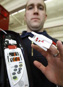
 AP Photo/Toby Talbot Montpelier, Vt., Lt. Jake Larrabee displays a RAD-57 in February after his department secured a grant for the devices. |
The detection of carbon monoxide poisoning is becoming increasingly important to prehospital providers. Invisible and odorless, CO fails to produce reliable signs or symptoms in poisoned patients that might alert health care providers or even patients themselves of significant poisoning. CO is the leading cause of poisoning deaths in every industrialized nation.
And while exact mechanisms are poorly understood, long term effects in survivors of CO poisoning include up to triple the incidence of cardiovascular events and deaths as well as disabling neurologic conditions. With increased awareness and availability of devices to measure CO in patients, the challenges of detecting CO poisoning should begin to decline. Prehospital providers using non-invasive CO screening devices are better prepared to assess and treat CO poisoned patients who might otherwise be missed.
Two technologies currently exist for field measurement of CO in people: CO-Oximetry and exhaled CO breath monitors. CO-Oximeters are pulse oximeters that utilize multiple wavelengths of light to detect not only oxygen saturated blood cells (oxyhemoglobin) but also carbon monoxide saturated blood cells (carboxyhemoglobin) and blood cells that have been chemically converted to methemoglobin. While many protocols exist for prehospital use and interpretation of CO levels, none have been published for prehospital evaluation of methemoglobin (METHb) levels.
Methemoglobin is formed when iron on the hemoglobin molecule is converted from a ferrous to a ferric state through a chemical reaction known as oxidation. METHb behaves in much the same manner as carboxyhemoglobin: it cannot carry oxygen and causes oxygen already bound to blood cells not to release itself into the body. The result is significant oxygen deficiency (hypoxia). Unlike CO poisoning, methemoglobinemia is relatively uncommon outside of medical settings. Most cases are caused by oxidizing drugs or chemicals. Worldwide, the leading cause of MET poisoning is the anti-malaria drug dapsone. While dapsone use is on the rise in the United States (for prevention of pneumocystis pneumonia in transplant patients on immune suppressants), the greatest cause of MET poisoning is benzocaine, a topical anesthetic agent used for numbing sensitive areas of the body. Absorption of benzocaine-containing sprays through mucous membranes or skin during intubations, endoscopies, bronchoscopies, and other procedures can induce significant MET poisoning.
Other acquired causes of MET poisoning include lidocaine and nitrates such as nitroglycerine, nitric oxide, or water sources contaminated by runoff containing nitrogen based fertilizers. The gastrointestinal tract of newborns may promote growth of gram negative organisms that induce MET poisoning usually associated with prolonged (> 7 days) infantile diarrhea. Methemoglobinemia can also occur as an inherited condition.
Assessment of methemoglobinemia
Like CO, the signs and symptoms of MET poisoning are vague and appear flu-like. Following an endoscopic procedure, patients may mistakenly believe they contracted a cold or flu virus while in the procedure lab. Symptoms in such cases are more likely related to methemoglobinemia than viral illness. Assessment of methemoglobinemia is by percentage of hemoglobin found in the ferric state. Treatment is typically reserved for symptomatic patients with MET levels above 30 percent. Untreated, methemoglobin has a half life (time for half the MET to be eliminated from the body) of roughly 55 minutes. Methemoglobin is continuously produced and eliminated in the body, and normal levels should not exceed 2 – 3 percent. Unlike CO poisoned patients, MET poisoning typically results in cyanosis at levels above 15 percent. Levels above 60 percent are lethal.
Treatment for methemoglobinemia is intravenous methylene blue dosed at 1 to 2 milligrams per kilogram of patient weight given over 5 minutes. Improvement is typically seen within 30 minutes. An additional dose of 1 milligram per kilo can be given at 30 minutes if improvement is not evident. Pediatric dosing (recommended only for children over 6 years old) is 1 milligram per kilo given intravenously over 5 minutes. Administration of methylene blue will completely skew readings and measurements of pulse oximeters, CO-Oximeters, and many lab instruments for at least 30 minutes.
Only one prehospital device is presently capable of measuring both CO and MET: the RAD-57cm™ (Masimo Corporation, Irvine, Calif. – Masimo.com). While some question the value of having technology capable of measuring a relatively uncommon prehospital condition, there is considerable benefit in using MET to confirm the accuracy of CO readings. A phenomenon common to most CO-Oximeters (including both blood gas analyzers and pulse co-oximetric devices) is a tendency to report falsely elevated CO readings in the presence of significant methemoglobinemia. While there are no published data on the accuracy of the RAD-57 in MET poisoned patients, the technology employed is strikingly similar to CO-Oximetric blood gas analyzers. As such, a user should be slightly suspicious of elevated CO readings when MET levels exceed 3 percent and be downright distrustful of reported CO values when MET levels exceed 5 percent.
Here’s an example of using MET to confirm CO. You are called to a 45-year-old female complaining of flu-like symptoms. The first response engine company reports a SpCO level of 45 percent using obtained with their RAD-57. They are not able to detect CO in the residence using a four gas meter. The hyperbaric chamber is 150 miles away, necessitating air medical transport. You assess the patient using a RAD-57cm and obtain a SpCO level of 42 percent and a SpMET level of 10 percent. Further questioning reveals the patient had a bronchoscopy yesterday at an outpatient surgery center and has been ill since her return home. You decide the reported CO level is likely to be falsely elevated because of methemoglobinemia and elect not to fly the patient for hyperbaric treatment. On arrival at the emergency department, a methemoglobin level of 10 percent is confirmed and the actual CO level is determined to be 2 percent. A wise use of technology? You betcha.
The practice of screening for CO and MET can be incorporated into EMS protocols using the following caveats:
Screening for CO and MET using a RAD-57cm ™ CO-Oximeter | ||
Rules: | 1. Transport any symptomatic patient regardless of readings | |
2. For any SpCO reading > 5%, always check SpMET% | ||
SpCO% Interpretation (carboxyhemoglobin): | ||
0 – 5% | Normal in non-smokers | |
5 – 10% | Normal in smokers | |
10 – 15% | (In any patient) assess for s/s, treat with high flow O2 if present | |
> 15% |
| |
> 30%, or | Consider immediate transport to closest hyperbaric treatment facility | |
SpMET% Interpretation (methemoglobin): | ||
0 – 3% | Normal in all patients | |
> 3% | SpCO level may be suspect – interpret cautiously | |
> 5% | Do not base treatment/transport decisions on SpCO readings | |
> 10% |
| |
> 30% |
| |
References:
Ash-Bernal R, Wise R, Wright SM. Acquired methemoglobinemia: A retrospective series of 138 cases at 2 teaching hospitals. Medicine. 83(5):265-273.
Betten DP, Vohra RB, Cook MD, Matteucci MJ, Clark RF. Antidote use in the critically ill poisoned patient. Journal of Intensive Care Medicine. 21(5):255-277.
Barker SJ, Tremper KK. The effect of carbon monoxide inhalation on pulse oximetry and transcutaneous P(O2). Anesthesiology. 66(5):677-679.
Barker SJ, Tremper KK, Hyatt J. Effects of methemoglobinemia on pulse oximetry and mixed venous oximetry. Anesthesiology. 70(1):112-117
Barker SJ, Curry J, Redford D, Morgan S. Measurement of carboxyhemoglobin and methemoglobin by pulse oximetry: A human volunteer study. Anesthesiology. 105(5):892-897.
Hampson NB, Weaver LK. Noninvasive CO measurement by first responders. A suggested management algorithm. Journal of Emergency Medical Services. 31(5): S10-12.
Henry CR, Satran D, Lindgren B, Adkinson C, Nicholson C, Henry T. Myocardial injury and long-term mortality following moderate to severe carbon monoxide poisoning. JAMA. 295(4):398-402.
Longo LD. The biological effects of carbon monoxide on the pregnant woman, fetus, and newborn infant. American Journal of Obstetrics and Gynecology. 129(1): 69-103.
MICROMEDEX Healthcare Series: Thomson Micromedex, Greenwood Village, Colorado (accessed September, 2008).
Weaver LK, Hopkins RO, Chan KJ, Churchill S, Elliott G, Clemmer TP, Orme JF, Thomas FO, Morris AH. Hyperbaric oxygen for acute carbon monoxide poisoning. New England Journal of Medicine. 347(14):1057-1067.
