

Practice ECG 2 (click for a larger image):
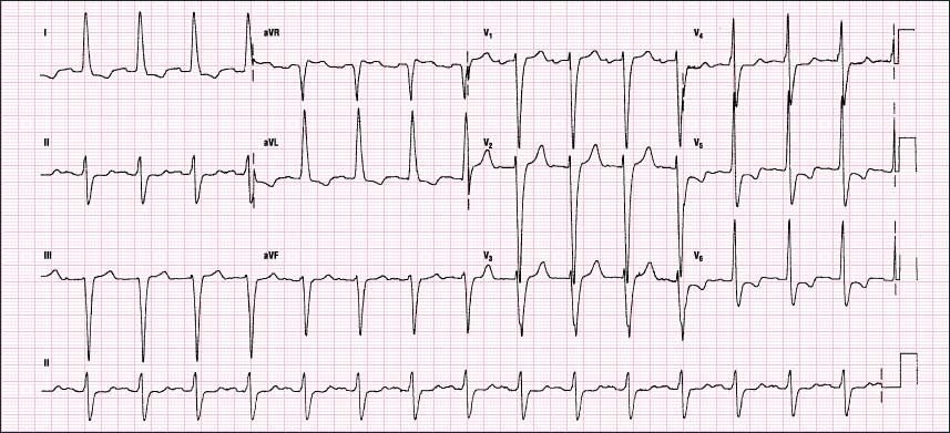 |
The P waves are hard to find, but they are there. Look at leads I and II. Use a magnifying glass if you have to, and take a good look at them. You will notice that they are wide-exactly 0.12 seconds-and notched, with the notches 0.04 seconds or more apart. This is an example of P-mitrale.
The P wave in V1 is also biphasic and meets LAE criteria. Once again note that the dimensions of the wave are small on the height scale.
The ECG also has an LBBB configuration. The axis is in the left direction in this case. Remember that an LBBB can have a normal, right, or left axis. The axis in an LBBB pattern depends on the pathway that the impulse takes as it is conducted through the ventricles. Let’s look at how this happens. In an LBBB pattern, the right bundle is working normally, and the right side of the heart depolarizes first. The left ventricle then depolarizes by direct, cell-to-cell transmission of the impulse.
This transmission will eventually touch the Purkinje system of either the left anterior fascicle or the left posterior fascicle. If it touches the left anterior fascicle first, or both divisions simultaneously, the axis is normal or right. This is because the superior and lateral walls of the ventricle are innervated first and then the rest of the ventricle is depolarized. This causes a more prolonged vector in the right or normal direction; hence, the axis shift. If, however, the left posterior fascicle is touched first, the axis will be directed to the left. In this case, the posterior and inferior aspects of the left ventricle are depolarized first, with the resultant prolonged vector occurring in a leftward direction as the lateral and superior walls are depolarized.
The T waves in V5 and V6 are concordant and possibly represent ischemia. Why is there a terminal S wave in V6 when there should not be one? Most probably, this is due to incorrect lead placement. When the electrodes are placed incorrectly in an anterior or superior location, the lead will show an S wave. Look at lead I to see the true morphology if the lead had been placed correctly. Remember that in the limb leads, placement of the electrodes is not as critical. For example, the right arm lead can be placed in the right wrist or shoulder and still appear the same. Therefore, small changes in position will not affect the limb leads as they do the precordials.
